Resorptive Lesions, Feline
Feline Odontoclastic Resorption Lesions (FORLs)
By Dr. Karen Burgess
Feline odontoclastic resorption lesions (FORLs) are a common (20-75%) dental disease in cats over 4 years of age. In this disease, cells known as odontoclasts, which originate in the bone marrow or spleen, migrate and attach to the external surface of the tooth root (portion of the tooth within the tooth socket) and resorb (i.e. destroy) the root surface. The odontoclasts are cells that are normally involved in the process of turning over “baby” teeth before the permanent teeth erupt. Although not known why, these cells remain active in the adult cat.
Over time, the root(s) of the teeth are completely destroyed, and in the latter stages of the disease, only the crown (portion of the tooth above the gumline) or portions of the crown remain. In many cats, the end-stage affected tooth is observed as missing.
FORLs were previously and incorrectly referred to as a feline cavity. We now know that cavities and FORLs are distinctly different diseases. Cavities are caused by bacteria, and FORLs, although their true cause is unknown, are not a bacterial disease. Many potential causes for this disease have been investigated, but to date, the true cause of the disease remains elusive, and is one of the current “hot” topics of research in the field of veterinary dentistry.
What are the symptoms of FORLs?
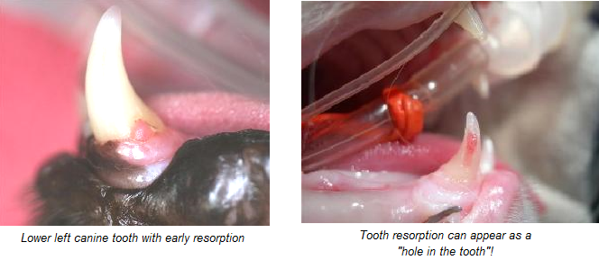
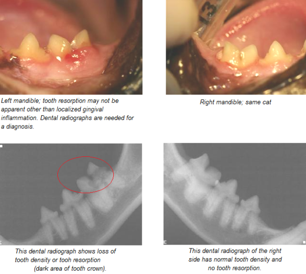
Cats with FORLs are often identified by the chief symptom of teeth “chattering,” sensitivity upon eating/chewing (i.e., dropping food or preferentially chewing on one side of the mouth). Patient’s suffering with FORLs may also salivate profusely. This suggests that there is significant oral pain. On examination, the veterinarian will identify either missing teeth or teeth where portions of the tooth crown are missing. In areas where portions of the crown are missing, the gums in the area are usually observed to cover the missing area, and a red spot is noticed on the crown. The teeth with early FORLs cannot be identified on gross examination, because the disease is localized to the root surface, and can only be documented by radiographs.
Symptomatic cats are usually those that have teeth with partially missing tooth crowns, and where the disease process has moved beyond the root surface. In other cats, despite the severe gross clinical appearance of the lesion, the cat remains unaffected in its behavior pattern: eating, gaining weight and content.
NOTE: Cats are very careful not to demonstrate pain. Signs of pain can be very subtle with these cats. You might notice more calculus (tartar) in specific areas, gingival inflammation (possibly the only sign), increased salivation or changes in food preferences. Owners typically fail to realize their cat is painful until after they experience behavioral changes (happier and more playful cats) subsequent to treatment for these resorbing teeth.
What tests are needed?
Only the end-stage lesions involving the tooth crown can be identified readily on clinical examination, the remainder of lesions must be diagnosed by quick and efficient dental x-rays which is available at Healthy Paws Animal Hospital. Due to a high percentage of cats affected by this disease, cats over the age of 4 are recommended to have dental x-rays as a screening test for the disease when having their teeth cleaned.
What treatment is needed?
FORLs are believed to be a painful disease in the cat, and cats with documented disease should be treated. The primary treatment for this disease is extraction of the affected teeth. When FORLs were believed to be similar to cavities, the lesions or defects in the crown were filled, similar to human cavities. As the disease was further investigated, and follow-up was performed on teeth that had been filled, it became clear despite our best efforts the filled teeth continued to resorb.
Extraction or crown amputation with intentional root retention, are the only currently accepted methods of therapy. The latter is a procedure where the crown of the affected tooth is removed with a bur; leaving the resorbing roots buried in the bone to continue resorbing to completion. The crown amputation procedure alleviates the clinical signs of disease because the exposed and sensitive portion of the tooth is removed. This procedure, however, is limited to affected teeth that have been appropriately x-rayed and have severe tooth root resorption. Teeth with severe root resorption are difficult to impossible to extract, and severe damage to the surrounding bone may result from overzealous attempts at extraction.
Prognosis
The prognosis following extraction or crown amputation of affected teeth is good, but affected cats will always have a predisposition to the development of additional lesions. Follow-up visits on an annual basis are recommended for FORL screening.
Preventative Care
It is helpful to brush your cat’s teeth daily or three times weekly at a minimum. Dental products are available that have been noted to reduce plaque and tartar. Products exhibiting the VOHC label have proven their efficacy and should be purchased over products that do not carry the VOHC label. VOHC products include treats, food, and water additives. Ask a Healthy Paws employee for a list of approved products.