Category Archives: Ears
Ear Meds- How To
How to Administer Ear Medication
By Dr. Karen Burgess
Understanding the Ear
- Always remember that the ear is a delicate structure. When inflamed or infected it may be very painful.

- The ear is really just a canal or tube lined by skin. If a pet has underlying skin disease it is common to also have ear issues and it is fairly uncommon for a pet to just “get an ear infection”. Instead it is often a sign of underlying skin disease or systemic allergies.
- The anatomy of some breeds can affect the ear’s ability to stay healthy. Whether it is a too narrow passage or an ear with excessive secretions these are typically lifelong issues.
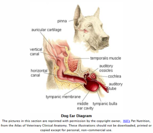
- The ear canal is L-shaped with a vertical and horizontal portion. The vertical portion is visible to the naked eye, the horizontal portion is visible only via a veterinarian’s otoscope. The tympanic membrane (TM) lies at the end of the horizontal canal. Often a pet owner may see a normal appearing vertical canal while the otoscope may reveal a completely occluded horizontal canal. To address ear disease both the vertical and horizontal portion of the ear must be treated while protecting the TM. The TM or eardrum can be damaged by disease, medications, or objects placed into the ear (i.e. Qtips). While the eardrum can heal, damage can also potentially affect hearing longterm.
Medicating 101
- If you feel you are not able to safely treat your pet’s ears, do not proceed. Ear cleaning can be scary and painful which could potentially make a normally docile animal become aggressive.
- Before starting, gather supplies: ear medication, towels, treats, old clothing.
- Location, location, location-find an area where your pet can be confined. An elevated surface or corner of a room often works well. Having an extra pair of hands is often helpful. For smaller animals a towel can be used to “burrito” wrap and better control the process.
Process
- Find an appropriate area to work in. Flooring with good grip or the ability to have your pet sit with their bottom against a corner or wall may be helpful.
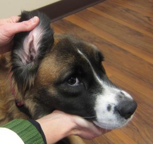
- Hold the ear flap up vertically with your non-dominant had allowing visualization of the ear canal.

- Holding medication in dominant hand, place the opening of the ear medication (often a spigot or long slender dropper) over the opening into the ear canal and gently squeeze instilling ointment or liquid into canal.

- Hold the ear flap (also called the pinna) closed like a resealable bag. Massage the base of the ear briefly as long as not too uncomfortable for pet.
- Allow pet to shake head, medication may come out but some will stay in. Wipe inner ear flap if necessary to clean off any residual debris.
- Give treats and verbal praise throughout process.
Ear Mites..
Ear Mites
By Dr. Karen Burgess
What exactly are ear mites?
Ear mites are microscopic bugs that physically resemble ticks and reside in the ear canal and skin of cats and dogs. The adult ear mite may be visible using a magnifying glass as a small moving white dot. Ear mites produce debris and discharge in the ear canal that can easily be misdiagnosed as a yeast or bacterial ear infection if not looking specifically for ear mites.
What are the signs of ear mite infection?
Ear mites live and breed in the ear canal, specifically on the surface of the skin. During this process the mites feed on oils and ear wax and subsequently cause inflammation in the ear canal producing a black discharge and general ear inflammation. Infected pets will often have painful itchy ears, head shaking, crusting on the skin around the ears, and notable ear discharge. The adult ear mite can travel outside of the ear canal to the surrounding skin and fur making systemic treatment preferable to treating just the ears.
How do dogs and cats get ear mites?
Ear mites are highly contagious and transmitted by direct contact. Cats contract ear mites more commonly then dogs. It is not uncommon to diagnose ear mites in pets coming from shelter or group housing situations.
How are ear mites diagnosed?
A microscopic examination of debris from the ear will typically show actual ear mites or their eggs.
How are ear mites treated?
There are a variety of treatments for ear mites. Topical ear drops have been a common ear mite treatment with some products available even over the counter. A disadvantage of topical treatment is that they do not kill ear mite eggs and thus involve twice daily ear drops for a minimum of three weeks. For some pets this can be uncomfortable and difficult to accomplish. Tresaderm is a prescription topical that does kill eggs and only requires twice daily treatment for two weeks. Neither topical product addresses mites that have migrated out of the ear canal. Alternatively the topical product Revolution can be applied twice (one month between doses) to the skin between the shoulder blades. This treatment effectively kills mites in the ears and on the skin. Another treatment option is the dewormer ivermectin which can be given as an injection or orally. This treatment is considered off-label meaning it is not appropriate for all pets. All pets in the household (including ferrets and rabbits) should be treated simultaneously. Pet bedding should also be washed.
Can humans contract ear mites?
Ear mites are not considered contagious to humans.
Otitis
Otitis (Ear Inflammation)
By Dr. Karen Burgess
What is otitis?
Otitis is better known as an ear infection. Otitis externa would be infection of the external ear, media the middle ear, and interna the inner ear (where the hearing apparatus is located).
What are the signs of otitis?
Dogs and cats with otitis may present with ear redness, discharge or odor. Some may rub or scratch at their ears. Owners may notice pain or reluctance to having the head and ears pet.
How do dogs and cats get otitis?
There are a variety of reasons pets get ear infections. Bacteria and yeast are common causes, but are typically considered secondary to some other pre-existing condition. The wax, moisture, and oils found in the ear canal contribute to problems by “feeding” the infection. Anatomically, some breeds with narrow ear canals, floppy ears, or a tendency toward excessive fur in their ears may have more frequent ear infections. Ear mites or foreign bodies can lead to secondary infections of the ear. Pets that have had recurrent or untreated previous ear infections may develop scar tissue making future episodes of otitis more likely. For pets experiencing repeated infections, underlying allergies (food or inhalational) are common and should be considered as a primary cause.
How is otitis diagnosed?
Physical examination of the ear canal will show discharge and inflammation associated with otitis externa and media. Otitis interna requires advanced imaging and anesthesia for diagnosis. Cell samples are taken from the ear canal to examine microscopically for the presence of bacteria and/or yeast. In some cases a culture of the ear may be recommended to determine specific bacterial presence. Debris can also be evaluated for evidence of ear mites and their eggs. If underlying allergies are suspected a food allergy trial, antihistamines, or referral to a dermatologist for skin testing may be recommended.
What is the treatment for otitis?
Ear infection treatment varies depending on severity. Cleaning of the ear canal may be recommended, but in some cases is considered too painful and irritating initially. Ear medications are often instilled into the ear by owners at home once to twice daily. Rechecking the ear for complete response in one week may be recommended. In some cases wax impregnated medication may be instilled in the veterinary office. For ears experiencing chronic disease surgery may eventually be indicated.
What are possible complications from otitis?
Vestibular disease (similar to vertigo in humans), ear hematomas (broken blood vessel in the ear flap), and hearing loss are all possible sequel to otitis.
How can otitis be prevented?
Prevention of ear infections is most successful when underlying disease is treated and predisposing conditions controlled. For some pets, preventative ear cleansings may be beneficial on a regular schedule or after swimming.
Ear Hematoma
Ear Hematoma
By Dr. Karen Burgess
What is an ear or aural hematoma?
An ear hematoma is a typically non-painful pocket of blood that develops in the ear flap or pinna after rupture of a blood vessel. Similar to a “blood blister” the cartilage of the ear flap contains the blood creating a swelling on the ear flap that often resembles a pillow or water balloon.
How do dogs and cats get ear hematomas?
Ear hematomas occur after a blood vessel ruptures in the ear flap. This often occurs after a traumatic episode such as rough play, bite injury, or aggressive flapping of the ears. It is not uncommon for pets with ear infections or severe ear inflammation to develop an ear hematoma.
How is an ear hematoma diagnosed?
Ear hematomas are diagnosed by visual examination or aspiration by a veterinarian. Ear cytology may also be recommended to look for an underlying ear infection.
How are ear hematomas treated?
There are a variety of treatments that have been used over the years for ear hematomas. In the end, this is not typically a life threatening issue and more of an aesthetic concern. It is important to know that regardless of treatment method there is likely to be some degree of scarring. The scarring may be more noticeable in pets with upright ears. For this reason, some may opt to not pursue any treatment, letting the body reabsorb the blood and essentially scar. This in turn avoids the expense, discomfort, and follow up involved with surgical repair. Lack of treatment may cause more extensive scarring similar to a “cauliflower ear”. Surgical treatment can range from incising the hematoma and stitching it flat to placing a cannula into the hematoma for several weeks. If surgery is performed it is extremely important to protect the surgery site either via and Elizabethean collar or a bandage. There are some veterinarians that have also had success using steroid injections to treat ear hematomas. Some might wonder why the ear hematoma cannot just be drained. Unfortunately this is usually only a short lived solution with the hematoma quickly refilling.

